Looking at the full scope of PAD: Cases, techniques, and economic considerations for complex disease.

< Table of Contents

Case Report: Treating a Long Chronic Total Occlusion in a Patient With CLI

A personalized treatment approach to a challenging case.

BY ERIK WEEKMAN, BS; VINAYAK SUBRAMANIAN, BS; IVY SMITH, BS; AND GEORGE L. ADAMS, MD, MHS, FACC, FSCAI

Critical limb ischemia (CLI) is associated with very high morbidity and mortality and is caused by inadequate arterial blood flow to the extremities. CLI is often associated with complex atherosclerotic disease including long chronic total occlusions (CTOs). Treating CTOs is technically challenging due to a variety of proximal and distal cap configurations and mixed lesion morphology.1,2 These lesions are often long and cross multiple vascular beds. The following case presented such a challenge; a personalized approach is described for access, crossing, and treatment to provide the best outcome for this patient.

Figure 1. Flush occlusion of the SFA (A). Distal reconstitution of the peroneal artery (B).

Figure 1. Flush occlusion of the SFA (A). Distal reconstitution of the peroneal artery (B).

CASE PRESENTATION

A 64-year-old woman with a history of hypertension, dyslipidemia, stroke, diabetes, and peripheral artery disease (a stent had previously been placed in the distal left superficial femoral artery [SFA]) presented with nonhealing wounds of the plantar aspect of the left lower extremity. Angiographically, the patient did not have significant disease in the distal aorta, bilateral iliac arteries, or common femoral artery (CFA) systems. In the left lower extremity, the SFA was occluded at its ostium and reconstituted at the proximal peroneal artery (Figure 1). This was the sole vessel to the foot; it reconstituted the posterior tibial artery at the ankle and supplied the plantar region of the foot, where the wounds resided.

INTERVENTION

A 6-F, 45-cm sheath was placed to the level of the left CFA from a contralateral retrograde CFA access. The proximal cap of the left SFA CTO was probed with an 18-g CTO wire but was unsuccessful in entering the proximal segment of the SFA. Therefore, direct angiographic access of the left popliteal stent was performed with an 18-gauge needle and a stiff TERUMO GLIDEWIRE® Guidewire (Figure 2A). The stiff TERUMO GLIDEWIRE Guidewire was supported with an 0.035-inch TERUMO NAVICROSS® Support Catheter and the CTO was successfully crossed within the true lumen and into the left CFA. An angled COOK® CXI® Support Catheter was then placed into the left CFA from the 6-F sheath, and the retrograde stiff TERUMO GLIDEWIRE Guidewire was exchanged for an 0.014-inch, 18-g–tip load COOK® APPROACH® CTO Microwire Guide. The wire was then maneuvered into the COOK CXI Support Catheter and externalized in a flossing fashion (Figure 2B). Laser atherectomy was then performed with a 2-mm SPECTRANETICS® TURBOELITE Laser Atherectomy Catheter at 50/40 rate/fluency and 60/60 rate/fluency from the ostium of the left SFA to the mid aspect of the distal SFA stent. Because this was restenotic plaque, there was high suspicion for the presence of thrombus in the plaque. Laser atherectomy was the preferred modality for treatment in this case due to its indication for use with in-stent restenotic plaque.3 This was followed by percutaneous transluminal angioplasty (PTA) of the SFA with a 4- X 200-mm balloon. On angiography, it was obvious that a large amount of thrombus was present at the ostium of the left SFA, so a 6- X 50-mm GORE® VIABAHN® Endoprosthesis was placed to prevent shifting of plaque to the lower extremity. The GORE VIABAHN Endoprosthesis was also used to ensure the stent was accurately placed in the SFA and prevent blocking the deep femoral profunda artery (Figure 3). This stent successfully trapped the thrombus and resulted in < 10% residual stenosis of the vessel. A second wire, an 18-g BOSTON SCIENTIFIC® VICTORY Guidewire, was then extended past the retrograde access site and internal tamponade was performed with a 6- X 40-mm balloon for 3 minutes as the TERUMO NAVICROSS Support Catheter was removed from the distal SFA stent (Figure 4).

Figure 2. Angiography-guided (RAO 30º) access of the prior stent in the left SFA (A). Retrograde TERUMO GLIDEWIRE® Guidewire guided into angled COOK® CXI® Support Catheter to “floss” and externalize retrograde wire through the contralateral CFA access site (B).

Figure 2. Angiography-guided (RAO 30º) access of the prior stent in the left SFA (A). Retrograde TERUMO GLIDEWIRE® Guidewire guided into angled COOK® CXI® Support Catheter to “floss” and externalize retrograde wire through the contralateral CFA access site (B).

Figure 3. Preintervention SFA, homogenous plaque noted in the vessel (A). Postintervention SFA after placement of GORE® VIABAHN® Endoprosthesis (B). Soft plaque is successfully trapped by the stent.

Figure 3. Preintervention SFA, homogenous plaque noted in the vessel (A). Postintervention SFA after placement of GORE® VIABAHN® Endoprosthesis (B). Soft plaque is successfully trapped by the stent.

Figure 4. Internal tamponade of retrograde access site.

Figure 4. Internal tamponade of retrograde access site.

The popliteal and tibioperoneal trunk occlusion was then crossed with the BOSTON SCIENTIFIC VICTORY Guidewire and exchanged for an 0.017/0.014-inch CSI® VIPERWIRE Advance-CSI-Diamondback-Guide-Wire, and a small ABBOTT® EMBOSHIELD NAV6 Embolic Protection Device was placed in the peroneal artery (Figure 5). The filter was placed to protect the sole vessel to the distal extremity and reduce the risk of distal embolization. PTA was performed with a 4- X 200-mm balloon. The filter was then removed. The intervention ultimately led to direct inline flow to the distal vascular bed (Figure 6).

Figure 5. ABBOTT® EMBOSHIELD NAV6 Embolic Protection System deployed in the distal peroneal prior to protect sole outflow vessel.

Figure 5. ABBOTT® EMBOSHIELD NAV6 Embolic Protection System deployed in the distal peroneal prior to protect sole outflow vessel.

Figure 6. Postintervention. Revascularized distal SFA— popliteal (A). Direct inline flow to distal vascular bed from the peroneal artery (B).

Figure 6. Postintervention. Revascularized distal SFA— popliteal (A). Direct inline flow to distal vascular bed from the peroneal artery (B).

DISCUSSION

CLI is a complex disease and treatment remains technically challenging. There have been significant technical advancements in the treatment of complex peripheral artery disease, and care must be individualized to meet the patient’s specific needs. The case described in this article illustrates the importance of understanding principles of access, crossing, and treatment and applying advanced techniques such as direct stent access to improve the chances of technical success. Furthermore, CTOs are a challenging entity and require an understanding of advanced crossing techniques such as retrograde crossing and flossing techniques. CTOs often have complex plaque morphology and are characterized by both homogenous and calcific plaque. In addition, in-stent restenotic disease is associated with the presence of thrombus, and the proper use of laser atherectomy to modify and treat the plaque is essential. Finally, it is important to realize that the distal lower extremity was only being perfused by one vessel, and protecting the vessel by trapping soft plaque with a covered stent and placing a filter is an important measure to prevent distal embolization and further complicating an already challenging case.

Ultimately, technical success in the treatment of complex peripheral artery disease is influenced by three tenants: (1) the skill and knowledge of the interventionalist, (2) patience during the case, and (3) the nuanced use of the tools available to overcome the many challenges commonly encountered in the treatment of CLI.

 < Previous Article                                        Next Article  >
 

  1. Saab F, Jaff MR, Diaz-Sandoval LJ, et al. Chronic total occlusion crossing approach based on plaque cap morphology: the CTOP classification. J Endovasc Ther. 2018;25:284–291.
  2. Torii S, Mustapha JA, Narula J, et al. Histopathologic characterization of peripheral arteries in subjects with abundant risk factors [published online December 6, 2018]. JACC Cardiovasc Imaging.
  3. Dippel EJ, Makam P, Kovach R, et al. Randomized controlled study of Excimer laser atherectomy for treatment of femoropopliteal in-stent restenosis. JACC Cardiovasc Interv. 2015;8:92-101.
GORE VIABAHN Endoprosthesis

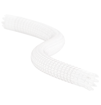
Proven across a broad range of complex cases, the versatility of the GORE® VIABAHN® Endoprosthesis with PROPATEN Bioactive Surface* enables you to deliver high patency and durable outcomes to minimize interventions for your patients.

Endovascular Today February 2019 Supplement

Endovascular Today February 2019 Supplement
Looking at the Full Scope of PAD


Download now

Erik Weekman, BS
North Carolina State University
Raleigh, North Carolina

Disclosures: None.
 

Vinayak Subramanian, BS
UNC School of Medicine
Chapel Hill, North Carolina

Disclosures: None.
 

Ivy Smith, BS
UNC REX Healthcare
Raleigh, North Carolina

Disclosures: None.
 

George L. Adams, MD, MHS, FACC, FSCAI
Associate Professor
UNC School of Medicine
Chapel Hill, North Carolina
Director
Cardiovascular and Peripheral Vascular Research
UNC REX Healthcare
Raleigh, North Carolina

Disclosures: Consultant to Gore & Associates; Spectranetics, a Philips Company; and Cook Medical.