Looking at the full scope of PAD: Cases, techniques, and economic considerations for complex disease.

< Table of Contents

Case Report: Aortoiliac Occlusion in a Patient With Severe Claudication

Management with covered balloon-expandable stents provides another option for complex disease.

BY EHRIN J. ARMSTRONG, MD, MSc

A 64-year-old man with a past medical history of coronary artery disease and prior smoking presented with severe bilateral lower extremity claudication on exertion. Ankle-brachial indices (ABIs) demonstrated a right ABI of 0.75 and a left ABI of 0.85. The patient was initially treated with a walking program and optimal medical therapy, but he continued to experience severe lifestylelimiting claudication. Therefore, he was referred for lower extremity angiography and possible endovascular intervention.

Treatment

Aortoiliac angiography (Figure 1A) demonstrated a small- to moderatecaliber aorta with a focal 70% to 80% stenosis at the origin of the left common iliac artery and an occlusion of the right common iliac artery just after its origin. The distal right common iliac artery reconstituted just above the level of the right internal iliac artery. The ongoing bilateral external iliac and common femoral arteries were relatively free of disease and the patient did not have any obstructive infrainguinal disease.

The decision was made to proceed with endovascular repair of the iliac arteries. Bilateral 7-F sheaths were placed in the common femoral arteries under ultrasound guidance. The right common iliac artery occlusion was crossed from a retrograde approach with a straight stiff TERUMO GLIDEWIRE® Guidewire and the occlusion was then predilated with a 4-mm balloon (Figure 1B). A 7- X 59-mm GORE® VIABAHN® VBX Balloon Expandable Endoprosthesis (VBX Stent Graft) was deployed in the right common iliac artery and a 7- X 39-mm VBX Stent Graft was deployed to the stenosis at the origin of the left common iliac artery (Figure 1C). These stents were simultaneously postdilated to 9 mm.


Figure 1. Baseline angiography revealed a small-caliber aorta with an occluded right common iliac artery (left arrows) and a high-grade stenosis of the left common iliac artery at its origin (right arrow). The ongoing external iliac arteries and bilateral internal iliac arteries were patent (A). The right common iliac artery occlusion was successfully crossed and then predilated with a 4-mm balloon (arrow) (B). After predilation, a 7- X 59-mm GORE® VIABAHN® VBX Balloon Expandable Endoprothesis (VBX Stent Gr

Figure 1. Baseline angiography revealed a small-caliber aorta with an occluded right common iliac artery (left arrows) and a high-grade stenosis of the left common iliac artery at its origin (right arrow). The ongoing external iliac arteries and bilateral internal iliac arteries were patent (A). The right common iliac artery occlusion was successfully crossed and then predilated with a 4-mm balloon (arrow) (B). After predilation, a 7- X 59-mm GORE® VIABAHN® VBX Balloon Expandable Endoprothesis (VBX Stent Graft) was deployed to the right common iliac artery, and a 7- X 39-mm VBX Stent Graft was deployed to the left common iliac artery (arrows) (C).

After stent deployment in the bilateral common iliac arteries, a residual stenosis remained in the distal right common iliac artery just proximal to the origin of the right internal iliac artery (Figure 2A). Therefore, an additional 7- X 29-mm VBX Stent Graft was deployed at nominal pressure and then postdilated to 9 mm in the right common iliac artery, while taking care to ensure that this stent did not impinge on the origin of the right internal iliac artery (Figure 2B). Final angiography revealed excellent stent expansion and no residual stenosis (Figure 2C). Bilateral ABBOTT® PERCLOSE PROGLIDE® Suture-Mediated Closure Systems were deployed for hemostasis, and the patient was discharged home later the same day on a regimen including aspirin at 81 mg daily and clopidogrel at 75 mg daily. At 1-month follow-up, duplex ultrasound revealed patency of the common iliac arteries, and the patient reported that he did not experience any claudication and was now able to walk more than 2 miles per day.


Figure 2. After initial stent deployment, there was excellent stent expansion in the bilateral common iliac arteries, but a residual stenosis at the distal right common iliac artery near the origin of the right internal iliac artery (arrows) (A). A 7- X 29-mm VBX Stent Graft was deployed (arrow), while taking care to not occlude the internal iliac artery (B). Final angiography revealed excellent stent expansion and flow throughout the iliac arteries (C).

Figure 2. After initial stent deployment, there was excellent stent expansion in the bilateral common iliac arteries, but a residual stenosis at the distal right common iliac artery near the origin of the right internal iliac artery (arrows) (A). A 7- X 29-mm VBX Stent Graft was deployed (arrow), while taking care to not occlude the internal iliac artery (B). Final angiography revealed excellent stent expansion and flow throughout the iliac arteries (C).

Discussion

Covered balloon-expandable stents have a number of advantages in the treatment of complex aortoililac disease. The randomized COBEST trial has demonstrated improved long-term patency of aortoiliac lesions when covered balloon-expandable stents are utilized instead of noncovered stents.1 In the VBX FLEX Study, primary patency rates for the VBX Stent Graft were 96.9% at 9 months.2 In addition, recently presented 24-month data on the VBX Stent Graft showed a 93.1% freedom from target lesion revascularization.3

Covered balloon-expandable stents have procedural advantages compared with noncovered stents. For example, the use of a covered stent can prevent any perforation that may occur during aortoiliac intervention.4 Although perforation is a rare occurrence, it can be life threatening if not treated immediately. By using a covered stent in areas of high calcification or after subintimal crossing of an iliac chronic total occlusion, the likelihood of any significant perforation occurring is reduced or eliminated. Covered stents can also exclude plaque characteristics, including thrombus or calcification, that may lead to embolization during a procedure. For all these reasons, covered balloon-expandable stents may be preferred for the treatment of complex aortoiliac lesions.

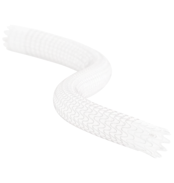
The VBX Stent Graft has a number of features that improve on previously available covered balloon-expandable stents, including an expanded size matrix, increased flexibility (data on file; W. L. Gore & Associates, Inc.; Flagstaff, AZ), and predictable deployment to the target lesion with excellent accuracy and minimal foreshortening.2 In this case, the common iliac artery stents were deployed at 7 mm but were easily postdilated to 9 mm with no evidence of significant shortening. The residual stenosis in the distal right common iliac artery was also easily treated with an additional VBX Stent Graft, which could be predictably deployed without altering flow to the right internal iliac artery. Both of these features of the VBX Stent Graft helped achieve an optimal procedural result, and the patient has done well during follow-up.

Conclusion

Covered balloon-expandable stents have furthered the treatment options for patients with complex aortoiliac disease by providing improved patency and an optimal safety profile. As techniques continue to evolve, almost all patients with aortoiliac disease can be treated with endovascular techniques.

 < Previous Article                                        Next Article  >
 

  1. Mwipatayi BP, Sharma S, Daneshmand A, et al. Durability of the balloon-expandable covered versus bare-metal stents in the Covered versus Balloon Expandable Stent Trial (COBEST) for the treatment of aortoiliac occlusive disease. J Vasc Surg. 2016;64:83-94.e1.
  2. Bismuth J, Gray BH, Holden A, et al. Pivotal study of a next-generation balloon-expandable stent-graft for treatment of iliac occlusive disease. J Endovasc Ther. 2017;24:629-637.
  3. Holden A. Comparison of four balloon expandable covered stents for the treatment of aorto-iliac occlusive lesions: which, where and when? Presented at: the 45th Annual VEITHsymposium; November 13–17, 2018; New York, NY.
  4. Aggarwal V, Waldo SW, Armstrong EJ. Endovascular revascularization for aortoiliac atherosclerotic disease. Vasc Health Risk Manag. 2016;12:117-127.
GORE VIABAHN VBX Balloon Expandable Endoprosthesis
Designed to expand to every demand, the GORE® VIABAHN® VBX Balloon Expandable Endoprosthesis (VBX Stent Graft) offers proven procedural success and long-term outcomes through flexibility, strength and accuracy to treat aortoiliac occlusive disease.

Endovascular Today February 2019 Supplement

Endovascular Today February 2019 Supplement
Looking at the Full Scope of PAD


Download now

Ehrin J. Armstrong, MD, MSc
Associate Professor of Cardiology
University of Colorado
Aurora, Colorado

Disclosures: Consultant to Abbott; Boston Scientific Corporation; Cardiovascular Systems, Inc.; Gore & Associates; Medtronic; and Philips.